Maths 'can help implants fit better'
Vienna, May 3, 2012
Individuals with implants may soon be able to feel the benefit of basic scientific knowledge in their own bodies, a research project by the Austrian Science Fund FWF said.
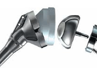
The project demonstrated how 3D models and special mathematical methods could be used to improve the design and integration of implants in the body on a patient-specific basis.
Data was gathered from computer and magnetic resonance tomography and used to generate 3D models specifically for shoulder joints and their replacements. The data was analysed in a procedure known as the finite element method, and possible individual optimisations were calculated. The project exemplifies the acute benefit of research findings from the Translational Research Programme, which ended at the close of the first quarter of 2012.
Basic research forms the foundation for future applications, as illustrated by programmes like the Translational Research Programme. This programme, which the Austrian Science Fund (FWF) conducted on behalf of the country´s Federal Ministry for Transport, Innovation and Technology (BMVIT), ran until early 2012 and served to accelerate the transfer of basic knowledge into practical applications: Applications which, first and foremost, improve the quality of people´s lives, in addition to creating economic value.
SHOULDER TO SHOULDER - MATHEMATICS & MEDICINE: This project brought together basic scientific knowledge from the areas of mathematics, medicine and computer science with the aim of optimising replacement shoulder joints individually (patient-specific). Headed by Dr Karl Entacher from Salzburg University of Applied Sciences and Dr Peter Schuller-Götzburg from the Paracelsus Medical University in Salzburg, the project initially computed human shoulder joint models and then used them as the basis for the analytical simulation of varying load conditions.
The team commenced by using imaging techniques to create the computer models. To this effect, computer tomography was used to build up images of human shoulder joints on a layer-by-layer basis.
As Dr. Entacher explains: "Modern tomography techniques allow us to create images of an entire shoulder joint layer-by-layer, and the layer thicknesses that we can achieve today make excellent resolution possible. We were able to use this image data to create computer-generated 3D models of each patient´s individual shoulder joint, forming the basis for our subsequent analysis." - TradeArabia News Service


